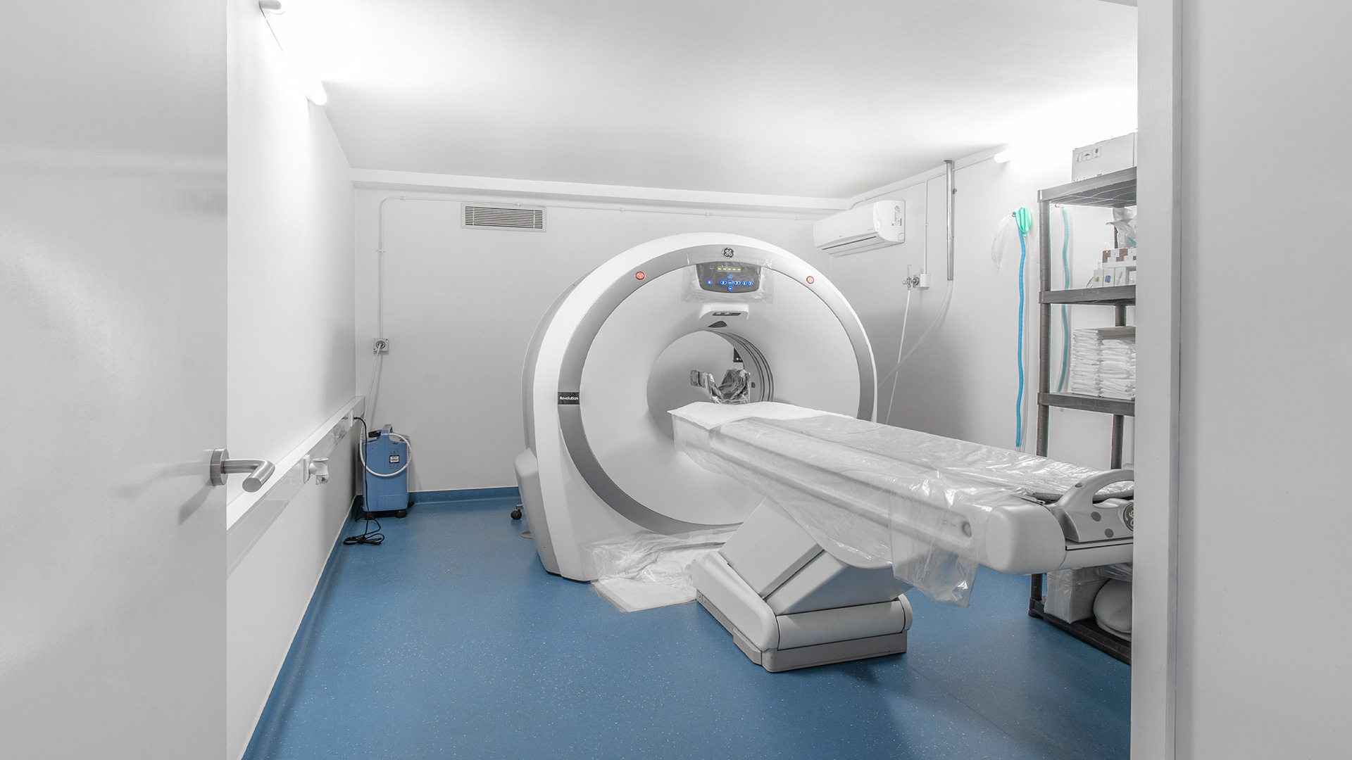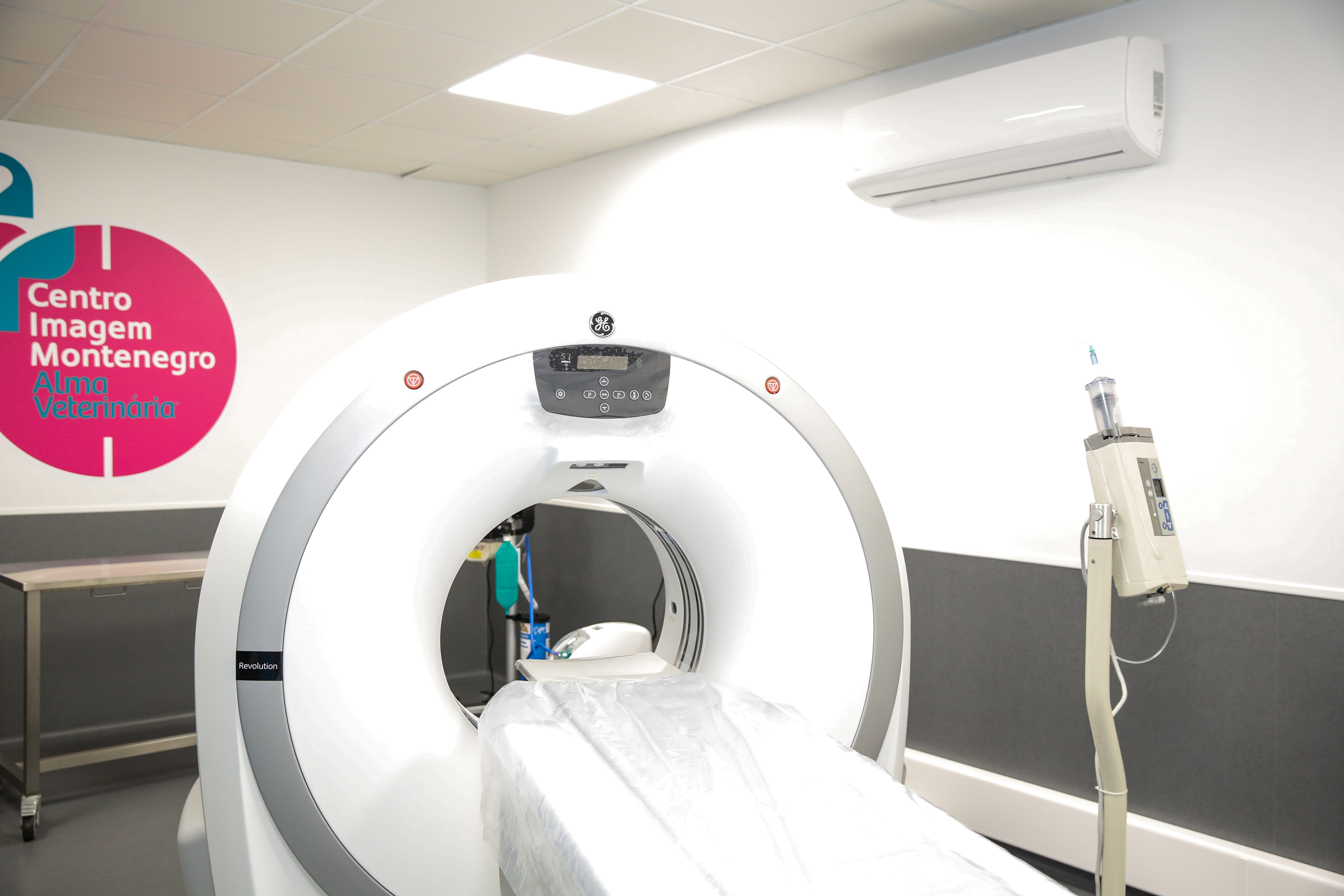
The Computed Tomography (CT) is an advanced imagiology diagnostic exam, which is non invasive, and uses radiation X to adquire images of the animal body in a transversal plain. The machine send rays of radiation X on the body of the animal that are then detected in the detectors that are rotating all together in 360 degrees around the animal’s area we want to study. The CT allows us to study several different anatomic structures, avoiding the overlapping of organs or bones that happen in the convetional x-ray
CT scan are done in several different parts of the body and used for large aray of pathologies, such as, congenital diseases, inflammatory and infectious diseases, neoplasic and traumatic diseases. Here are some examples of the most common studies we make:
– Cranium Studies: Studies of the upper respiratory system, teeth and mandibule diseases, external medium and internal ear, traumatic injuries and fracture studies.
– Thoracic Cavity: Lower respiratory system (Trachea and lung), thoracic chest, pleural space, esophagus and mediastinum structures
– Abdominal-pelvic Studies: Diseases related to the abdomen (Liver, Gastrointestinal system, urinary system, adrenal glands, liver, reproductive diseases)
– Vascular Studies: Angiography (With Automatic Contrast Media Injector)
– Axial and apendicular skeleton: Vertebral spine, thoracic and pelvic limb studies.